

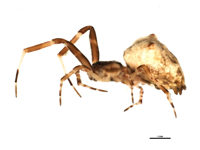
As the sun sets, the hackled orb-weaving spider Uloborus diversus goes to work. Slender and pale, with banded legs bearing a fine brush of comb-like hairs (Fig. 1), this spider gently hangs midair from a silk line. Thread by thread, it lays out the tapestry of its web, weaving a geometric pattern using a brain not much larger than a poppy seed. Each placement demands precision—knowing where it is in space, how taut each strand feels, the pull of the wind, and how the growing web unfolds around it. How can a relatively small brain perform such a feat? A new three-dimensional brain atlas offers insight into this question, revealing a potential hidden web-building architecture.
Gregory Artiushin, Abel Corver, and Andrew Gordus [1] created the new spider brain atlas by first dissecting out whole brains, chemically treating them to render the tissue transparent, and staining them with immunofluorescent markers. Each preparation was labeled for synapsin—a protein that localizes to presynaptic sites at which neurons communicate—and paired with additional stains for major neurotransmitters and neuropeptides. These included the fast transmitters acetylcholine and GABA, which mediate excitation and inhibition; amines like dopamine, serotonin, and octopamine, which shape arousal and motivation; and peptides such as Proctolin, Allatostatin A, CCAP, and FMRFamide, which modulate circuits involved in movement, feeding, and other behaviors [reviewed in 2]. The intact, cleared brains were then imaged with confocal microscopy, which optically sections tissue plane by plane without physical slicing. Data from several spiders were averaged together to generate a standardized 3D scaffold that served as the reference brain for later mapping.
The results were striking (and beautifully illustrated in the accompanying colorful figures). The arcuate body, previously recognized as a lobed but largely uniform midline structure, revealed far more internal complexity when viewed with multiple stains, including layered subdivisions and distinct zones of innervation. Adjacent to it, the team identified a newly described “tonsillar neuropil” (named for the way it encircles the esophagus as it passes through the brain)—a dense, fan-shaped region with organized serotonergic and octopaminergic input. They also mapped a protocerebral bridge and commissure, each with characteristic neurochemical signatures. Taken together, these features bear a strong resemblance to components of the insect central complex (reproduced in Fig. 2), a structure well known for its role in path integration and navigation [reviewed in 3].

The mushroom bodies, by contrast, lit up bright with synapsin but showed surprisingly little evidence of neuromodulator labeling. This pattern suggests dense synaptic activity without broad neuromodulatory input, the former of which is consistent with their putative role in processing vibratory feedback. For an orb weaver, such feedback is indispensable: vibrations tell the spider whether a thread is properly placed, whether silk is taut, or whether prey has struck the web. A previous landmark comparative study by Skye Long found robust mushroom bodies in both orb web-building spiders and highly visual hunters [4], suggesting an important role for these structures in vibratory and visual motion processing, respectively.
Taken together, these findings hint at a division of labor for web building: a central complex-like region for orienting in space, and mushroom bodies for integrating vibratory cues.
In my view, this study offers convincing evidence that spiders generally possess a central complex analog. Perhaps later studies will even find another anatomical ring-like structure that functions like a compass, whether in the arcuate body or across neuropils. That would not be surprising, given the shared ancestry of arthropods and the universal need to navigate space. In insects, the central complex is well established as a hub for orientation, while in vertebrates, hippocampal place cells provide a complementary system for encoding maps of space [reviewed in 5]. I speculate that spiders may combine aspects of both: orientation computed in the arcuate body and its central complex-like circuitry, with spatial maps and place-like coding emerging in the mushroom bodies.
It is important to note, however, that finer anatomical data (e.g., globuli cells and glomerular organization [6]) and genetic data (e.g., specific learning and memory proteins [7]) suggest that the mushroom bodies of spiders and insects derive from the same ancestral brain region. This ancestral region likely facilitated the processing of incoming chemical information from a dedicated olfactory appendage, such as antennae [4], which was lost in the lineage that gave rise to modern spiders. In insects, mushroom bodies are involved not only in olfactory processing but also in learning and multisensory integration, and they exhibit remarkable plasticity. Thus, if the mushroom bodies in U. diversus are embedded within a central complex–like aggregation of brain regions that support spatial behavior, this would more likely reflect the co-option of an existing brain region for new computations rather than the persistence of an ancestral, cryptic component of the central complex. That said, I am more convinced by the protocerebral bridge and tonsillar neuropil being ancestral components of a central complex. Figure 2 evokes an interesting question: to what extent are structural similarities a coincidence, from shared ancestry, or might they reflect a constraint of optimal computation?
The implications from this study extend beyond orb weavers. Jumping spiders, which do not spin webs but stalk prey visually and return to nests with remarkable spatial fidelity [8], may rely on similar central brain structures tuned to distant visual landmarks instead of vibrations.
I also want to highlight the pioneering work of Andrew Gordus and colleagues at Johns Hopkins, who approach the problem from behavior inward: characterizing structured web-building routines [9] and linking them to neural [1] and genetic [10] underpinnings. This kind of integrative effort is needed, not only for spiders but for a wide array of “non-model” organisms. By expanding beyond the usual laboratory animals, we can uncover both the common principles of neural computation and the unique strategies that have arisen through evolution.
This atlas provides a crucial framework, but the next step must involve combining quantitative behavior, connectomics, cell-type physiology, and transgenics to truly resolve how these circuits operate. Populations of cells that stain for the same neurotransmitter protein likely consist of many different cell types with distinct functions, and it’s hard to resolve the fine structure of brain regions with immunostaining alone. As neuroscience moves into the tool-rich and comparative era, spiders stand out as powerful models. This brain atlas is an essential first step—an anatomical map that reveals hidden structure and provides a scaffold for future discovery. With it, we are now positioned to ask deeper questions about how small nervous systems generate behaviors as intricate as web-weaving and navigation, and to ask further questions of how small brains give rise to complex behavior.
Disclosure: I served as a reviewer of the eLife publication discussed here.
References
[1] Gregory Artiushin, Abel Corver, Andrew Gordus (2025). A three-dimensional immunofluorescence atlas of the brain of the hackled-orb weaver spider, Uloborus diversus. eLife14:RP107732, https://doi.org/10.7554/eLife.107732.1
[2] Jékely, G. (2013). Global view of the evolution and diversity of metazoan neuropeptide signaling. Proceedings of the National Academy of Sciences, 110(21), 8702-8707.
[3] Pfeiffer, K., & Homberg, U. (2014). Organization and functional roles of the central complex in the insect brain. Annual review of entomology, 59(1), 165-184.
[4] Long, S. M. (2021). Variations on a theme: Morphological variation in the secondary eye visual pathway across the order of Araneae. Journal of Comparative Neurology, 529(2), 259-280.
[5] Leutgeb, S., Leutgeb, J. K., Moser, M. B., & Moser, E. I. (2005). Place cells, spatial maps and the population code for memory. Current opinion in neurobiology, 15(6), 738-746.
[6] Strausfeld, N. J., Hansen, L., Li, Y., Gomez, R. S., & Ito, K. (1998). Evolution, discovery, and interpretations of arthropod mushroom bodies. Learning & memory, 5(1), 11-37.
[7] Wolff, G. H., & Strausfeld, N. J. (2015). Genealogical correspondence of mushroom bodies across invertebrate phyla. Current Biology, 25(1), 38-44.
[8] Winsor, A. M., Remage-Healey, L., Hoy, R. R., & Jakob, E. M. (2024). Visual attention and processing in jumping spiders. Trends in Neurosciences, 47(1), 6-8.
[9] Corver, A., Wilkerson, N., Miller, J., & Gordus, A. (2021). Distinct movement patterns generate stages of spider web building. Current Biology, 31(22), 4983-4997.
[10] Miller, J., Zimin, A. V., & Gordus, A. (2023). Chromosome-level genome and the identification of sex chromosomes in Uloborus diversus. GigaScience, 12, giad002.
